Showing posts with label Pulmonary. Show all posts

21 y/o female presents with dysphagia and pleurisy after getting into an argument and yelling loudly at her boyfriend. What is the treatment based on her chest x-ray?

Click on image to enlarge.

Scroll down for answer


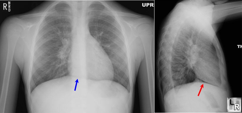
Pnuemomediastinum.  Notice the free air in the neck on the chest x-ray. 

Spontaneous, atraumatic, pneumomediastinum usually occurs when air leaks through small alveolar ruptures triggered by asthma or Valsalva maneuver and is a benign condition that resolves on its own in 2 to 15 days.  Treatment includes analgesia and avoidance of maneuvers that increase pulmonary pressure, ie. Valsalva.   Bronch is not indicated for screening of patients with isolated spontaneous pneumomediastinum.  An esophageal study (contrast esophagography or esophagoscopy) is not indicated unless there is clinical concern for esophageal rupture.  Rare complications of spontaneous pneumomediastinum that necessitate more aggressive care include tension pneumomediastinum and pneumopericardium.


Source 

Saadoon, A. and Janahi, I.  "Spontaneous pneumomediastinum in children and adolescents"  Up to Date.  May 2011.

What is the cause of this "white out" on the right side of the chest x-ray?

Pleural effusion.  Click image to enlarge.

CT of same patient demonstrating left pleural effusion.

A couple features suggest pleural effusion over other causes:
  • homogeneous opacity with no air bronchograms
  • concave upper surface producing a meniscus 
  • while not really notable with this pleural effusion, some large pleural effusions will displace the mediastinum towards the contralateral side 

    The "white out" is unlikely to be caused by lobar collapse because there is no evidence to suggest decreased lung volume (displacement of pulmonary fissure, elevation of hemidiaphragm, displacement of hila, displacement of mediastinum or compensatory overinflation of an adjacent lobe).   Collapse of each lobe has a characteristic radiographic appearance, click here to view.

    Pneumonia is also an unlikely cause.  For one, there is a second area of consolidation at the left lung base (also a pleural effusion).  While multifocal pneumonia does exist, non-infective pathology takes a higher place in the differential when there are multiple areas of consolidation.  Additionally, pneumonia generally has a more inhomogeneous opacification with air bronchograms and ill-defined margins. Click here to view a large pneumonia.


    Source

    Jenkins, P.  Making Sense of the Chest X-Ray: A Hands-On Guide, 1st ed.

    Lisle, D. Imaging for Students, 3rd ed.

    Schwartz, D.  Emergency Radiology: Case Studies

    Name seven causes of an elevated hemidiaphragm. What is the workup?

    Elevated right hemidiaphragm.

    7 causes of an elevated hemidiaphragm include:
    1. volume loss (lobar collapse, atelectasis)
    2. splinting 
    3. pleural disease (mass, effusion)
    4. diaphragmatic hernia 
    5. eventration
    6. phrenic nerve paralysis (most commonly from iatrogenic injury after cardiothoracic or cervical procedure; or from compression by malignancy)
    7. abdominal disease (abscess, dilated viscera)
    If the cause is not immediately obvious from initial history and physical, further workup - often as an outpatient - should be pursued and may include examination of diaphragmatic excursion under fluoroscopy and/or chest CT.  For patients found to have phrenic nerve paralysis, treatment is generally conservative unless the patient is symptomatic in which case operative management (diaphragmatic plication) is an option.


    Source

    ACS Surgery: Principles & Practice

      What is the cause of this chest x-ray opacity?

      click picture to enlarge

      CT of opacity.  Click picture to enlarge.


      Eventration of the diaphragm.  Caused by a thinned and weakened section of diaphragm - often congenital - permitting abdominal contents to bulge towards the thoracic cavity.  While eventration may cause respiratory distress in the newborn, adults are often asymptomatic.  The distinction between a localized eventration or a pathological mass is best made using CT or MRI.


      Source 

      Clinical Imaging: An Atlas of Differential Diagnosis.  5th ed.

      Adam: Grainger & Allison's Diagnostic Radiology, 5th ed.

      Which patients with hemoptysis should get an emergent chest CT?

      High risk patients (oncology, smoker) with minor hemoptysis and all patients with moderate to large hemoptysis should undergo plain chest x-ray followed by emergent chest CT. Hospital admission for observation and brochoscopy should also be considered.


      Source

      Marx: Rosen's Emergency Medicine, 7th ed.

      Identify the key steps to management of massive (> 500 cc in 24 hour period) hemoptysis in the emergency department.

      1. Asphyxiation, NOT blood loss, is generally the cause of death.  Intubate with size 8.0 or larger endotracheal tube.  Larger tube will facilitate bronchoscopy. 
      2. Obtain chest x-ray and place patient with bleeding site down, if identifiable, to prevent aspiration of blood into contralateral lung. If bleeding is from the left lung, consider advancing the endotracheal tube into the right main stem bronchus.
      3. Reverse coagulopathy and thrombocytopenia. 
      4. Consult pulmonary for bronch to identify and attempt to treat bleeding source.  
      5. Consider thoracic surgery and interventional radiology consultations. 

      Source

      Kruse: Saunders Manual of Critical Care, 1st ed.

      Ingbar, D.  "Massive hemoptysis: Initial management"  Up to Date.  2011 Jan.

      Identify the pathology on this AP and lateral chest x-ray of a patient who presents with chest pain s/p laprascopic appendectomy.

      Identify the pathology on this AP and Lateral chest x-ray of a patient who presents with chest pain s/p laprascopic appendectomy.

      Patient's chest x-ray

      Normal chest x-ray for comparison

      Pneumomediastinum.  Note the "continuous diaphragm sign" in the patient's chest x-ray caused by air collecting inferiorly between the heart and diaphragm.   Other signs of pneumomediastinum to look for, but which are not readily noted on this x-ray, include a thin layer of air adjacent to the heart or aorta separated from the lung by a fine line representing the displaced parietal pleura and air in the subcutaneous soft tissues of the chest and neck.

      Pneumomediastinum, along with pneumothorax and pneumopericardium, are potential complications of laparoscopic surgery which can occur if there are embryonic remnants constituting channels of communication between the peritoneal cavity and the pleural and pericardial sacs that open when intraperitoneal pressure increases during the creation of pneumoperitoneum.


      Source

      Schwartz, D.  Emergency Radiology: Case Studies.  2008. 

      Miller, R. et al.  Miller's Anesthesia, 7th ed.

      Image source, pneumomediastinum: http://www.learningradiology.com/archives05/COW%20132-Pneumomediastinum/pneumomedcorrect.htm

      Image source, normal chest x-ray: http://blog.dianahsieh.com/2010/07/chest-pain-in-80-year-old-woman.html

      This is the BEST vent lecture that I've ever heard

      "How to Dominate the Ventilator" by Scott Weingart

      Succinct
      Insightful
      Practical

      My management strategy has just evolved dramatically. Thanks Scott.


      Source

      http://learn.emcrit.org/uncurated-videos/

      What is the appropriate dose and duration of steroids to treat a COPD exacerbation?

      40 mg prednisone x 10 - 14 days.


      Source

      Niewoehner, D. "Outpatient Management of Severe COPD" N Engl J Med 2010; 362: 1407-16.

      2 y/o child chokes while eating peanuts but now in the ED is asymptomatic. Exam and chest x-ray (w/ lateral decub) normal. Bronch or not?

      Aspirated foreign bodies, particularly organic material, can cause pneumonia, lung abscess and empyema.

      Given the risks of retained aspirated foreign bodies and the small, albeit known risks of bronch (laryngeal lacerations, pneumomediastinum, pneumothorax, etc), a negative bronchoscopic evaluation rate of 10-15% is acceptable.

      Hence, the decision to bronch or not lies in the pre-test (bronch) probability of retained foreign body which can be assessed by the usual means (history, physical, chest x-ray [expiratory, decubitus]) as well as admission for short period of observation and/or expert consultation. CT and MRI are generally not part of the evaluation given the young age of the patients, time and expense involved and the need for sedation in a patient with potential pulmonary compromise.


      Source

      Taussig: Pediatric Respiratory Medicine, 2nd ed.

      What is the clinical significance of a chronic pulmonary embolism? How is it treated?

      A subset of patients with acute pulmonary emboli (PE), treated or not, go on to form chronic PE whereby the emboli get incorporated into the pulmonary arterial wall. The mechanism by which this occurs is unknown.

      The clinical significance of a chronic PE is that it is a potentially treatable cause of pulmonary hypertension either via pulmonary artery endarterctomy or the use of pulmonary vasodilator therapy.

      Chronic PEs can often be distinguished from acute PEs on CT angiogram with the former demonstrating tapering of pulmonary arteries, web-like strictures and other irregularities of the intimal surface and the later, acute PEs, demonstrating an abrupt vessel cutoff.


      Source

      Auger, W., et al. "Chronic Thromboembolic Pulmonary Hypertension" Clinics in Chest Medicine. 2007.

      Of the following clinical scenarios, choose the 2 where non-invasive ventilation (BIPAP, CPAP) has demonstrated the greatest efficacy.

      • neuromuscular disease
      • thoracic trauma
      • acute respiratory failure in asthma
      • end stage patients as palliative measure
      • extubation failure
      • copd exacerbations
      • cardiogenic pulmonary edema
      • obesity hypoventilation
      While non-invasive ventilation has been used and demonstrated some efficacy in each of the above clinical scenarios, there is greatest evidence for success in those with copd exacerbations and cardiogenic pulmonary edema.


      Source

      Nava, S. and Hill, N. "Non-invasive ventilation in acute respiratory failure" Lancet. 18 July 2009.

      Name two thoracic complications that may ensue from bowel perforation that results as a complication of colonoscopy.

      Depending on the site of colonic perforation, air from the insufflated bowel can enter into either the peritoneum or retroperitoneum.

      1. If air enters the peritoneum, gas can traverse from here through small fenestrations in the diaphragm and enter the pleural space along a pressure gradient causing a pneumothorax. Aside from minute diaphragmatic fenestrations, there is a subset of patients who have undiagnoosed diaphragmatic defects which allow the direct transmission of air into the thorax.

      2. If air enters the retroperitoneum a pneumomediastinum can ensue through direct communication.


      Source

      Zeno, B. et al. "Tension Pneumothorax Following Colonoscopy" Chest. Oct 2005. Supplement.

      What is the most common cause of pneumomediastinum ?

      Microscopic alveolar rupture.

      Clinical settings where this may occur:
      • deliberate alteration in breathing pattern: marijuana smoking and cocaine inhalation, pulmonary function testing, mountain climbing, wind instrument playing, yelling, shouting, singing
      • straining and other involuntary alterations in breathing pattern: childbirth, vomiting, seizures, coughing, sneezing, hi cupping, heavy lifting, athletic competition, straining at stool
      • external pressure changes: scuba diving, air travel, mechanical ventilation, Heimlich maneuver
      Other less common causes of pneumomediastinum include air escaping from the upper respiratory tract or GI tract.

      Source

      Mason: Murray & Nadel's Textbook of Respiratory Medicine, 4th ed.

      Should patients with a COPD exacerbation be treated with antibiotics?

      If mild (defined as not requiring mechanical ventilation and having only one of the three cardinal symptoms of increased dyspnea, sputum purulence or sputum production), NO.

      If moderate to severe (defined as requiring mechanical ventilation or having at least two of the three cardinal symptoms), YES.

      Which antibiotic?

      If no risk factors for complicated COPD (age < 65, < 3 exacerbations/year, no cardiac disease), treat with macrolide (azithromycin), cephalosporin, doxycycline or trimethrprim/sulfamethoxazole; if 1 or more risk factors and no risk factors for pseuduomonas, treat with fluoroquinolone (moxifloxacin, gemifloxacin, levofloxacin), amoxicillin/clavulanate or ceftriaxone; if 1 or more risk factors for complicated COPD and risk factors for pseudomonas, treat with levofloxacin, cefepime, ceftazidime or piperacillin-tazobactam.

      Bottom line, most folks that show up in the ED with a COPD exacerbation should recieve an antibiotic. Which type depends on their risk for complications and pseudomonas.

      Source

      Bartless, J. MD. Sethi, S. MD. "Diagnosis and treatment of infection in acute exacerbations of chronic obstructive pulomonary disease." Up to Date. Oct 2008.

      A patient with history of sarcoidosis presents with bronchitis symptoms. What's your recommended treatment?

      Common etiologies being common, a patient with these symptoms (cough, shortness of breath, chest discomfort and hemoptysis), even with history of sarcoidosis probably has a simple bronchitis and should be treated and evaluated as such. However, given that a sarcoid flare could present similarly if not exactly the same way, initiation of treatment with steroids to treat this should be considered at the time of ED evaluation or later if symptoms do not improve.

      Source

      King, T. MD. "Treatment of pulmonary sarcoidosis with glucocorticoids" Up to Date. Oct 2008.

      King, T. MD. "Clinical manifestations and diagnosis of sarcoidosis" Up to Date. Oct 2008.

      How long does it take for a tracheostomy tract to mature?


      • 5 days

      • when changing a tracheostomy tube before this time beware of false passage/lumen creation


      Source

      Roberts: Clinical Procedures in Emergency Medicine, 4th ed.

      When should embolectomy be considered for pulmonary embolus?


      • contraindications to thrombolytic therapy

      • hypotension despite maximal medical treatment for > 1 h


      Source

      Wood, Kenneth. "Major Pulmonary Embolism review of a Pathophysiologic Approach to the Golden Hour fo Hemodynamically Significant Pulmonary Embolism." Chest. V 121 Issue 3. March 2002.

      What are the hemodynamic changes that occur after intubation which may be deleterious to patients with a large pulmonary embolus?


      • sedative hypnotics used for intubation can blunt catecholamine surge on which patient is dependent for vasoconstriction and maintenance of venous return

      • hyperventilation can also decrease venous return

      • pulmonary vascular resistance may increase


      Despite these hemodynamic changes which may be deleterious to patients with large PE, these patients often require mechanical ventilation. Consider starting norepinephrine to counter some of these hemodynamic effects.

      Source

      Wood, Kenneth. "Major Pulmonary Embolism review of a Pathophysiologic Approach to the Golden Hour fo Hemodynamically Significant Pulmonary Embolism." Chest. V 121 Issue 3. March 2002.
      online dating chat rooms,free online dating chat,online dating chat tips,dating sites chat,chat avenue dating,online games dating,online chat singles,love dating online dating chat rooms,free online dating chat,online dating chat tips,dating sites chat,chat avenue dating,online games dating,online chat singles,love dating