Showing posts with label Radiology. Show all posts

Trick of the Trade: Ossification centers of the elbow


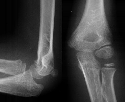
Fracture or a normal ossification center?

This is a common question heard when viewing an xray of a pediatric elbow. How do you remember the timing of normal ossification centers? FYI, the xray images above are normal and have no fractures.


Trick of the Trade:
The mnemonic CRITOE 1-3-5-7-9-11

  • Age 1 year:    Capitellum
  • Age 3 years:   Radial head
  • Age 5 years:   Internal (medial) epicondyle
  • Age 7 years:   Trochlea
  • Age 9 years:   Olecranon
  • Age 11 years: Extrenal (lateral) epicondyle


This great video is from a relatively new YouTube channel that I stumbled upon called "Radiology Channel", which is a Radiopaedia.org project featuring short polished presentations by Dr Andrew Dixon, Dr Frank Gaillard and Dr Jeremy Jones.

Mythbuster: Pediatric coin ingestion vs aspiration?


Is this coin in the esophagus or the trachea?

The classic teaching for the Boards exam is:

  • Esophageal coins appear in the coronal plane, as shown above.
  • Tracheal coins appear in the sagittal plane because of the cartilaginous tracheal rings.

Mythbuster: 
A case series of 8 pediatric patients were documented with coins positioned in a sagittal plane were actually in the esophagus! This data was collected over 15 years.

  • Age range 3-17 years old
  • Location of coin: 7/8 at level of aortic notch and 1/8 at distal esophagus 

The classic teaching likely still holds true most of the time (sagittal coin = tracheal foreign body), but don't rush to immediate judgment. Take a look at the lateral view, and see where the coin is located with respect to the trachea and airway. It may be more posterior, in which case, it's in the esophagus.

Thanks to Dr. Matt Anderson (Resident at Univ. of Wisconsin EM program) for the tip!


Reference

Schlesinger AE, Crowe JE. Sagittal orientation of ingested coins in the esophagus in children. AJR Am J Roentgenol. 2011 Mar;196(3):670-2. Pubmed .


Paucis Verbis: Diagnostic testing tips for acute abdominal pain


In the most recent EM Clinics of North America publication, Dr. Panebianco et al. discussed the evidence behind diagnostic tests for acute abdominal pain. There were some really great teaching points in this broad-reaching topic.

My favorite pearl: A 3-way acute abdominal series is too insensitive to rule-out any major acute causes of abdominal pain with confidence. So stop ordering them routinely. If you are worried about a perforated viscus, order an upright chest x-ray instead -- more accurate and less radiation.


You can download this PV card:  [MS Word] [PDF]

Reference
Panebianco NL, Jahnes K, Mills AM. Imaging and laboratory testing in acute abdominal pain. Emerg Med Clin N Amer . 2011;29(2):175-93. PMID: 21515175
.

Paucis Verbis: Blunt cerebrovascular injuries


In the setting of blunt trauma, it is easily to overlook a patient's risk for blunt cerebrovascular injuries (BCVI). These are injuries to the carotid and vertebral arteries. Often they are asymptomatic with the initial injury, but the goal is to detect them before they develop a delayed stroke.
  • Who are at risk for these injuries? 
  • What kind of imaging should I order to rule these injuries out? 
  • Do I really treat these patients with antithrombotic agents even in the setting of trauma to reduce the incidence of CVA?
FYI: A simple seat-belt sign along the neck does not warrant a CT angiogram. Patients with higher risk findings such as significant pain, tenderness, swelling, and/or a bruit probably need imaging.


You can download this PV card: [MS Word] [PDF]

Reference
Burlew CC, Biffl WL. Imaging for blunt carotid and vertebral artery injuries. Surgical Clinics of North America. 2011, 91(1), 217-31. PMID: 21184911
.

Paucis Verbis: Head CT clinical decision rules in trauma

The ideal clinical decision tool has a sensitivity and specificity of 100%.

You need a high sensitivity to be sure that your negative result indeed predicts a true negative. That means if your clinical decision tool suggests that you don't need to get a head CT, then your head CT would have been normal.

On the flip side, this realistically means there is a low-moderate specificity. That means a clinical decision tool with at least 1 positive criterion does not always mean that there will be an abnormal finding on head CT.

There are 3 major clinical decision rules that I've heard tossed around in the literature:
  • Canadian CT Head Rules (CCHR)
  • New Orleans Criteria (NOC)
  • National Emergency X-Radiography Utilization Study (NEXUS)-II 
There is no perfect tool.

Take a look at these decision rules and their inclusion criteria.
  • The CCHR included patients with GCS 13-15. The NOC initially enrolled only patients with a GCS of 15. 
  • All factor in age (≥65 years for CCHR and NEXUS-II; ≥60 years for NOC). 
  • Interestingly only the CCHR, for better or worse, take into account mechanism of injury. I'm not sure I would obtain a head CT on a pedestrian with a graze wound on the foot from a slow-moving vehicle.
Which do you use? I use a combination of all 3 and my clinical gestalt.


Trick of the Trade: Oblique CXR to look for pneumothorax

CT chest demonstrating a large left pneumothorax

Supine chest xrays have an extremely low sensitivity (12-24%) in detecting pneumothoraces. Because a pneumothorax layers anteriorly on an AP CXR film, the xray beam is perpendicular rather than tangential to the pneumothorax edge. This makes visualizing a small to moderate sized pneumothorax extremely difficult. So you are left to look for indirect signs such as a deep sulcus at the costophrenic angle or subcutaneous air. I'm often surprised at how large of a pneumothorax can be missed on CXR based on CT imaging.

What if you have an equivocal bedside ultrasound result in looking for a pneumothorax, and the patient is too unstable to go to CT?

Trick of the Trade:
Oblique CXR

Matsumoto et al in Annals of EM just reported this great tip. Position a film plate oblique to the patient's chest as shown in the above diagram from the article. "OPX" designates an Occult PneumothoraX. The x-ray beam is now more tangential to the edge of the anterior pneumothorax. This allow us to detect free air more easily.


Supine CXR, oblique view

Thanks to Dr. Fabio De Iaco and Dr. Gemma Morabito from MedEmIt for this CXR image.

Reference
Matsumoto S, Kishikawa M, Hayakawa K, Narumi A, Matsunami K, Kitano M. A method to detect occult pneumothorax with chest radiography. Ann Emerg Med. 2011, 57(4), 378-81. PMID: 20864214
.

Trick of the Trade: Finding the subtle pneumothorax


Can you see the pneumothorax?

Small pneumothoraces can be difficult to detect on chest xrays. Overlying ribs, other bony structures, and soft tissue can obscure subtle findings. For a patient at risk for a small pneumothorax, you can use your digital radiology PACS system to improve your ability to spot them.

Trick of the Trade: Invert digital image of chest film

Standard digital radiography systems allow the user to manipulate images on the screen. Specifically there is an “invert image” icon.

By clicking on the button, black images appear white, and white images appear black. Using this Invert feature, bones, soft tissue, and vascular markings will be black, and lung parenchyma will be white. The pleural line of a pneumothorax, now a dark line on a white background with this inverted setting, is often better visualized using this technique.

Check out the inverted image of the CXR showing a subtle right-sided pneumothorax (just inferior to the orange arrows). If only all xrays had these arrows...


EM Lightbox: Kidney stones


As most ED providers know, the term “kidney stone” is often inaccurate. Symptomatic patients typically present with stones not in the kidney itself but stuck at nature’s bottlenecks, i.e. the pelvic brim, ureteropelvic junction (UPJ), or most commonly the ureterovesical junction (UVJ)

Controversy exists regarding the optimal evaluation of nephroureterolithiasis. A non-contrast CT is often employed to diagnose stones, their size, location and associated ureterohydronephrosis, as well as to rule out other causes of pain. Finding the stone is crucial to prognosis, as stones that have passed into the bladder are passed nearly 100% of the time. In contrast, more proximally impacted stones may take longer to pass or may cause obstruction, infection, or intractable pain necessitating intervention.

The modern diagnostic approach is a prone noncontrast CT. Prone scanning helps delineate impacted UVJ stones from those layering posteriorly in the bladder near the UVJ. Pictured above is an impacted left UVJ stone, just adjacent to the bladder.

Pitfalls in locating stones in the distal ureters and bladder include phleboliths in pelvic veins and calcified seminal vesicles (seen above - calcified structure on the patient's right just posterior to the bladder).

References
Vaswani K, El-dieb A, Vitellas K, Bennett W, Bova J. Ureterolithiasis: classic and atypical findings on unenhanced helical computed tomography. Emergency Radiology 2002; 9: 60– 66.
_____________________________________________


This "EM Lightbox" case was authored by guest blogger, Dr. Eric Silman.

EM Lightbox: Lunate and perilunate dislocation

Lunate dislocation

Besides making frequent appearances on emergency medicine standardized exams, carpal dislocations, most commonly lunate and perilunate dislocations, present frequently to Emergency Departments. They are orthopedic emergencies and can be associated with significant complications.

Lunate dislocation (top image) occurs when the lunate is displaced and subluxed anteriorly. This often subluxes into the carpal tunnel causing an acute median neuropathy. It is seen as a “moon-“ or “saucer-shaped” bone flipped volarly, with the more distal capitate sliding proximally against the distal radius.

Perilunate dislocation

In a perilunate dislocation, the lunate-radius articulation is preserved and the capitate (as well as the entire distal carpal row with attached metacarpals) “jumps” dorsally “out of the saucer”.

Both of these carpal dissociations are often associated with scaphoid fracture and always portend significant ligamentous damage usually requiring operative repair.


Kienbock's disease

Another classic complication is Kienbock’s disease, or avascular necrosis of the lunate bone, which occurs with interruption of the bone’s tenuous blood supply, as in the scaphoid. Notice loss of bone density and height.

See prior post about normal wrist anatomy on xray.

References

Wheeless, C. “Perilunate dislocations.” Wheeless’ Textbook of Orthopaedics N.p., 30 Nov. 2008 Web. 17 Apr. 2010.

Voigt, C. Injury to the heel of the hand. Unfallchirurg. 2006; 109(4):313-22.

_____________________________________________


This "EM Lightbox" case was authored by guest blogger, Dr. Eric Silman.

EM Lightbox: Hemothorax on CXR

Upright CXR


Supine CXR


A hemothorax, a collection of blood in the pleural space, is relatively common in blunt or penetrating thoracic trauma and is an acute indication for pleural drainage.

Trauma patients are commonly initially evaluated with a supine chest x-ray. In traditional upright films, pleural blood layers inferiorly, obscuring the costophrenic angle laterally and/or the hemidiaphragm more medially. The top CXR shows a left hemothorax. Most studies agree that it takes somewhere between 200-500mL of fluid to blunt a hemidiaphragm.

In contrast, supine films may conceal sizeable hemothoraces. In supine films (bottom CXR), blood layers posteriorly, which causes generalized haziness of the affected hemithorax. Often the ipsilateral costophrenic angle and hemidiaphragm edges are preserved. In fact, up to 1 liter of pleural blood may go undetected on plain films.

With CT as the gold standard, bedside ultrasonoraphy by emergency physicians has been shown to be comparable in sensitivity to an initial supine chest x ray. This may expedite diagnosis.

References
Ma OJ, Mateer JR. Trauma ultrasound examination versus chest radiography in the detection of hemothorax. Ann Emerg Med. March 1997;29:312-316.
_____________________________________________


This "EM Lightbox" case was authored by guest blogger, Dr. Eric Silman.

EM Lightbox: Hyperdense MCA sign


Noncontrast CT of the brain is often used to evaluate patients with stroke-like symptoms, but is often normal or shows very subtle findings early in acute ischemic stroke.

One clue of acute ischemic stroke, shown above, is the "Hyperdense MCA Sign". The right Middle Cerebral Artery appears denser in its proximal course than the contralateral side. In this head CT, although it is not visualized in this CT slice, the contralateral MCA is normal in density.

The increased vessel attenuation on CT is thought to be due to thrombus within the MCA lumen. Studies have shown a high specificity but low sensitivity of this sign as well as poorer prognosis in patients who present with stroke and a hyperdense MCA sign. A false positive hyperdense MCA sign may be seen with increase hematocrit and vascular calcification.

References
Jha B, Kothari M. Pearls & Oysters: Hyperdense or pseudohyperdense MCA sign: A Damocles sword? Neurology 2009; 72: e116-e117.

Pettiti N. The hyperdense middle cerebral artery sign. Radiology 1998; 208:687–688.

_____________________________________________


This "EM Lightbox" case was authored by guest blogger, Dr. Eric Silman.

EM Lightbox: ACL rupture


The X-ray above shows two of the most common plain films signs of ACL rupture:
  • Avulsion of the lateral tibial plateau
  • Avulsion of the anterior tibial spine
The lateral tibial plateau avulsion, or Segond Fragment, is seen in greater than 75% of ACL tears. It has been shown to result from varus stress on the knee with internal rotation of the tibia, causing avulsion of bone at the insertion of the lateral joint capsule. Avulsion of the anterior tibial spine results from excessive tension on the ACL is usually accompanies rupture.

In acute knee trauma with pain and swelling, it is often difficult for emergency physicians to perform ligamentous examinations. These plain film hints can aid in the early diagnosis and prompt follow-up of ACL injuries.

References
Stallenberg, B. Fracture of the posterior aspect of the lateral tibial plateau: radiographic sign of anterior cruciate ligament tear. Radiology. 1993 Jun; 187(3): 821-5.

Wheeless, C. “Evaluation of the ACL tear.” Wheeless’ Textbook of Orthopaedics N.p., 30 Nov. 2008 Web. 17 Apr. 2010.
_____________________________________________


This "EM Lightbox" case was authored by guest blogger, Dr. Eric Silman.

Paucis Verbis card: Ottawa knee, ankle, and foot rules

Often times, I get called to triage to help decide whether a patient should be sent to Radiology for an initial x-ray after injuring their knee, ankle, and/or foot. After teaching one of the nurses about the Ottawa rules, she taped a list of these rules on the triage wall.

This installment of the Paucis Verbis (In a Few Words) e-card series reviews Ottawa Knee, Ankle, and Foot Rules.


Feel free to download this card and print on a 4'' x 6'' index card.

Paucis Verbis card: Ankle fractures

¿Hablas Inglés?
你讲英语?
هل تتحدث الانكليزية

Ankle fractures are a common injury diagnosed in the Emergency Department. Being able to speak Ortho-ese (i.e. the language of orthopedists) is invaluable in consulting the orthopedist over the phone. One ankle fracture classification system that our orthopedists like to use is the Lauge-Hansen system.

In this installment of the Paucis Verbis (In a Few Words) e-card series, the topic is the Lauge-Hansen classification of ankle fractures.


Feel free to download this card and print on a 4'' x 6'' index card.
[MS Word] [PDF]

Beware the hidden tibia plateau fracture

Find the fracture in this patient with blunt knee trauma.

As a general rule, plain films are insensitive in ruling out orthopedic injuries. One particularly tricky area is the knee. This 2-view knee series above is normal.

Did you know that the sensitivity of picking up knee fractures is as low as 79% with a 2-view series and 85% with a 4-view series? With the advent of CT imaging, more and more subtle fractures are being found.

Here is the same patient with 2 oblique views:


Although oblique views add more sensitivity in detecting fractures, these are also normal. However in the CT reformats of this patient's knee, the fracture becomes obvious - a depressed lateral tibial plateau fracture.


Beware the "invisible" tibial plateau fracture on plain films!

CT image the patient's knee especially if unable to bear weight after significant blunt trauma, despite plain films.

A radiology pearl: A subtle orthopedic diagnosis

A man recently presents with knee pain after pivoting and torquing his knee while falling. He complains of concurrent mild ankle pain. He presents with this tib-fib xray.


Realizing that a proximal fibular fracture can present concurrently with a medial malleolus fracture or deltoid ligament rupture, we obtained xrays of the ankle. We were looking for a Maisonneuve fracture.

Do you see an ankle injury?


Answer
  • There was no medial malleolus fracture.
  • There was no obvious deltoid ligament rupture, because the ankle mortise appears normal.
  • Always be sure to look at the lateral ankle view. You need to check for a posterior malleolus fracture.
  • This patient indeed had a Maisonneuve fracture, which was splinted and referred to orthopedics for operative repair.

Question: Have you had any interesting radiology findings on your ED shift?

Topics in Emergency Medicine CME Course


"Topics in Emergency Medicine" is one of two national CME courses that our UCSF department hosts annually, in addition to "High Risk Emergency Medicine" every May. This week Topics is being held in the fancy-schmancy Sir Francis Drake Hotel in San Francisco. It has an amazing line-up of speakers, most of whom are from UCSF and San Francisco General Hospital (SFGH).

I just gave a lecture yesterday on High Risk Back Pain, which focuses on key history and physical exam elements, imaging options, and advanced concepts in the 4 red flags of back pain (spinal fracture, cauda equina syndrome, spinal infection, and vertebral malignancy). My talk incorporates Flash-based modules, such as on the topic of reading thoracolumbar films. Basically, you should use an ABCS approach:
  • A- Alignment
  • B- Bone
  • C- Cartilaginous (i.e. disk space height)
  • S- Soft tissue


Thoracic spine
  • A- Anterior and posterior vertebral line
  • B- Vertebral body, pedicle, lamina, facet jt, spinous process
  • C- Intervertebral disk space
  • S - Paraspinous stripe


Lumbar spine
  • A- Anterior and posterior vertebral line
  • B- Vertebral body, pedicle, lamina, facet jt, spinous process
  • C- Intervertebral disk space
  • S - Psoas shadow


Article Review: Literature review of computer animations for medical education


"Animation is a series of images designed so that each image appears an alteration of the previous one, and in which the sequence of images is determined by either the designer or the user."

I came across this article evaluating the effectiveness of computer animation in learning, after having build an EM radiology animation module in a previous blog post. While I think it's cool, it would be nice to know if this approach to learning is better than showing just static images. From an instructor's standpoint, still images would be much easier to create.

There were many fascinating concepts in this article. For me, there were 4 key take-home points from:
  1. There are very few (and often conflicting) studies evaluating the effectiveness of animation vs static images in medical education.
  2. Allowing learner control of the animation pace improves the effectiveness of the animation.
  3. Building shorter (more digestible) animation pieces improves the effectiveness of the animation.
  4. There is a "cognitive load theory" which attempts to explain how people learn. The authors describe how animation fits into this framework.


Cognitive Load Theory Humans have a "working memory" center, which processes new information. Some of this information is stored in "long-term memory". There are 2 major types of information, or "cognitive loads".
  • Germane load: Information that contributes to learning.
  • Extraneous cognitive load: Information that takes up cognitive "space" in your working memory but does NOT add to effective learning.
Extraneous cognitive load (a.k.a white noise) wouldn't be a problem for learning except that our working memory has only a limited capacity for information. This contrasts long-term memory, which has no limits. So the goal behind teaching should be to maximize germane and minimize extraneous cognitive loads.

Cognitive Theory on Multimedia Learning
This theory builds on the "cognitive load theory" concept. It is known that people process information using visual and auditory pathways. Learning is improved if both pathways are engaged. That means both images and spoken words should be used (keeping in mind to minimize the extraneous cognitive load) to optimize teaching and learning.


So bringing this back to animation, what should we keep in mind if building animations for education?
Animations are not always superior to still images. Ask yourself, will the animation be more distracting than useful? Will this be considered a germane of extraneous cognitive load? Preliminary studies showed that building user-triggered controls, where the learner can control the animation pace or flow, improves learning comprehension.

Table 2 poses some interesting research questions, if you are interested in pursuing research in this area.
Click to enlarge table.


Reference:
Ruiz JG, Cook DA, Levinson AJ. Computer animations in medical education: a critical literature review. Med Educ 2009;43:838-46.
New EM radiology teaching idea: Wrist dislocation


What do you think is the best way to teach EM radiology?

Think outside the box.
Think beyond textbooks.
Think beyond static webpages.

I'm toying with the idea of using interactive Flash elements to teach radiology for emergency physicians. It's time intensive to build these modules, but the pay-off may be worth it. What do you think?

I've incorporated this approach to teaching radiology in a few of my past lectures, but I've never done so online. Here is my debut of a Flash-based teaching approach to reading plain films of the wrist.

Roll your mouse over the words to highlight the bones.

Image set #1: Normal xray of the wrist (AP and lateral) with key identifiable structures


Image set #2: Perilunate dislocation
For all plain films of the wrist, be sure to look at the lateral view for the 4-bone alignment (distal radius, lunate, capitate, 3rd metacarpal). Oftentimes, dislocations such as this perilunate dislocation are missed.


Image set #3: Lunate dislocation
Just yesterday, a Chicago Bears player Brian Urlacher sustained a lunate dislocation, which requires operative repair. Notice that the lunate appears abnormally as a triangle-shape on the AP view, instead of the usual cuboid shape. Furthermore, the lunate has "spilled" volarly on the lateral view. Both perilunate and lunate dislocations place the patient at risk for a median nerve injury.


online dating chat rooms,free online dating chat,online dating chat tips,dating sites chat,chat avenue dating,online games dating,online chat singles,love dating online dating chat rooms,free online dating chat,online dating chat tips,dating sites chat,chat avenue dating,online games dating,online chat singles,love dating