Showing posts with label ecg. Show all posts

Trick of the Trade: Speed up ECG paper rate to differentiate tachycardias


Undifferentiated tachycardias, especially when the rate is extremely fast, make it difficult to see anything other than the QRS complexes! Is there a P or flutter wave?

Trick of the Trade:
Double the ECG paper speed to 50 mm/sec

Standard ECG machines run at 25 mm/sec. If you double the paper output speed, subtle ECG findings hidden in the tracings become more evident. Imagine the ECG tracing as a string and that you are pulling on both ends. Everything, including the QRS complex and intervals, gets wider.

What are your experiences with this?

Atrial flutter at 150 bpm:
  • Standard rate 25 mm/sec 

  • Faster rate 50 mm/sec (red arrows are flutter waves)


Thanks to Dr. Amal Mattu for sharing his ECGs with me from his University of Maryland ECG archives. If you haven't heard of this amazing ECG video series, you should definitely check it out.

Below is the video teaching this point within the topic of narrow-complex regular tachycardias. This trick is seen at the 15:00 minute mark.


For another example, check out Dr. John Larkin's blog ECG of the Week post on a pediatric SVT rhythm at 300 bpm with no P waves seen at 50 mm/sec.


Reference
Accardi AJ, Miller R, Holmes JF. Enhanced diagnosis of narrow complex tachycardias with increased electrocardiograph speed. J Emerg Med. 2002 Feb;22(2):123-6. Pubmed .

Gaspar JL, Body R. Best evidence topic report. Differential diagnosis of narrow complex tachycardias by increasing electrocardiograph speed. Emerg Med J. 2005 Oct;22(10):730-2. Pubmed Free PDF

Paucis Verbis: Electrolytes and ECG changes


The electrocardiogram can pick up all sorts of electrolyte abnormalities. The most common abnormalities revolve around high and low levels of potassium and calcium. Magnesium derangements typically have nonspecific findings. How do you keep things straight? To make things more complicated, multiple electrolyte derangements can occur at the same time, making ECG interpretation challenging.

Thanks to Dr. Steve Field (Resident at Resurrection EM program) for the idea and card. Also take a look at Life In the Fast Lane's extensive review of ECG abnormalities and their causes.


Feel free to download this card and print on a 4'' x 6'' index card.
See other Paucis Verbis cards.


Reference

Diercks DB, Shumaik GM, Harrigan RA, Brady WJ, Chan TC. Electrocardiographic manifestations: electrolyte abnormalities. J Emerg Med. 2004 Aug;27(2):153-60. Pubmed .


Paucis Verbis: Blunt cardiac injury

From www.ctsnet.org

Do you always get a troponin for patients who sustain blunt chest trauma?

Hopefully your answer is no. Of note, it is also NOT indicated as a screening test for those in whom you suspect a blunt cardiac injury (BCI). It can be normal in the setting of arrhythmias and it can be falsely elevated in the setting of catecholamine release or reperfusion injury from hypovolemic shock.

The initial screening test should include an EKG and a FAST ultrasound exam. If you have abnormal EKG findings, then a troponin is warranted (in addition to hospital admission).

Below summarizes a suggested algorithm from the recent EM Clinics of North America publication series. Definitive statements are challenging because there is no gold standard to diagnose BCI.


Feel free to download this card and print on a 4'' x 6'' index card.

See other Paucis Verbis cards.


Reference

Bernardin B, Troquet JM. Initial management and resuscitation of severe chest trauma. Emerg Med Clin North Am. 2012 May;30(2):377-400. Pubmed .

Paucis Verbis: The aVR lead on EKG

What lead is the most overlooked on the EKG? 
aVR


Lead aVR can provide some unique insight into 5 different conditions:
  1. Acute MI 
  2. Pericarditis 
  3. Tricyclic antidepressant (TCA) and TCA-like overdose 
  4. AVRT in narrow complex tachycardias 
  5. Differentiating VT from SVT with aberrancy in wide complex tachycardias by using the Vereckei criteria (possibly better than Brugada criteria)
It turns out that aVR provides a lot of great information!


You can download this PV card:  [MS Word] [PDF]
References
  • Kireyev D, Arkhipov MV, Zador ST, Paris JA, Boden WE. Clinical utility of aVR-The neglected electrocardiographic lead. Ann Noninvasive Electrocardiol. 2010 Apr;15(2):175-80. .
  • Riera AR, Ferreira C, Ferreira Filho C, Dubner S, Barbosa Barros R, Femenía F, Baranchuk A. Clinical value of lead aVR. Ann Noninvasive Electrocardiol. 2011 Jul;16(3):295-302. .
  • Vereckei A, Duray G, Szénási G, Altemose GT, Miller JM. New algorithm using only lead aVR for differential diagnosis of wide QRS complex tachycardia. Heart Rhythm. 2008 Jan;5(1):89-98. .
  • Williamson K, Mattu A, Plautz CU, Binder A, Brady WJ. Electrocardiographic applications of lead aVR. Am J Emerg Med. 2006 Nov;24(7):864-74. .

Trick of the trade: Quieting the shaky EKG tracing


A patient with Parkinson's disease presents with chest pain to your ED. Her tremors prevent you from getting a good quality EKG because of the movement artifact.

How can you eliminate this artifact? (No cheating with rocuronium.)


Trick of the Trade
Have the patient sit on his/her hands.

Although I haven't tried this myself, this trick apparently works for people with upper extremity tremors and movement disorders. Has anyone used this trick? It's been referenced in: Burdick E350 Operating Manual. Milton, Wis: Siemens Burdick, Inc; 1990.

Thanks to Brooke, RN for this tip, and Alex for being a hand-sitting model!

Paucis Verbis: Brugada syndrome

Brugada, Brugada, Brugada

You always hear about it when working up syncope and sudden cardiac arrest in young patients, but it's so easy to forget what it looks like on EKG. We so rarely see it... or DO we?!

This Paucis Verbis card on Brugada Syndrome is to help emblazon these EKG tracings in our mind, so that we don't miss the subtle findings which place a patient at risk for sudden cardiac death. Pay special attention to Type 1, which is most specific for Brugada Syndrome.


References
Antzelevitch C et al. Brugada Syndrome: Report of the Second Consensus Conference: Endorsed by the Heart Rhythm Society and the European Heart Rhythm Association. Circulation, 2005; 111(5): 659-70. DOI: 10.1161/01.CIR.0000152479.54298.51

Free PDF article downloadable from Circulation.

Paucis Verbis: AMI and EKG Geography

(click to see enlarged view) 

Sometimes a picture is worth MORE than a 1000 words. Such is the case of the above illustration that I saw on the Life In The Fast Lane blog. When I first saw it, I knew that I immediately had to find out who made the graphic. It turns out it is the multitalented Dr. Tor Ercleve, who is an emergency physician at Sir Charles Gairdner Hospital and an established medical illustrator.

This graphic demonstrates the EKG findings for the various types of acute MI's as broken down by coronary vascular anatomy (right coronary artery, left circumflex artery, left anterior descending artery). This detailed illustration won't be readable in print form but is great in digital format on your mobile device.

Thanks, Tor!


Paucis Verbis: Right and posterior EKG leads


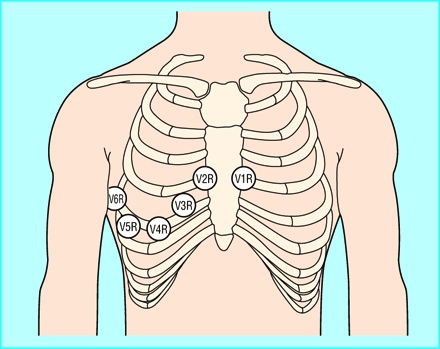
A standard 12-lead EKG can be very telling for patients with chest pain or shortness of breath. A right ventricular (RV) and posterior wall infarct, however, can present very subtly. You can obtain special right-sided (V1R-V6R) and posterior leads (V7-V9), if you are concerned.

What are the indications for obtaining right-sided and posterior EKG leads?


Addendum 3/11/11:
Right sided EKG leads (V1R-V6R) are positioned in a mirror image fashion from the standard 12-lead precordial leads.


Posterior EKG leads (V7-V9) are applied by moving V4-V6 in the posterior positions.

Feel free to download this card and print on a 4'' x 6'' index card.
Thanks to Caitlin for suggesting this topic!

Trick of the Trade: Getting an EKG on Chewbacca


Patients with a hairy chest may require little patches of hair to be shaved when applying EKG leads. This allows the leads to stick firmly to the chest. Loose leads will result in either an artifactual signal or no signal at all on the EKG machine.

How can you obtain an EKG without shaving little patches on the patient's chest?

Trick of the Trade: 
Cover EKG leads with damp gauze 

Water maximes the contact surface area between the EKG lead and the patient's skin. The water easily conducts the cardiac electrical signal.

Generously soak gauze with water.


Apply gauze over each EKG lead where it contacts hairy skin. 

Thanks to Dr. Gemma Morabito from Rome, Italy for telling me about this trick. Keep a lookout for more of Dr. Morabito's tricks on her Italian EM website, MedEmIt (http://www.medicinadurgenza.com/). 

Reference:

Article review: EKG interpretation by the computer

What is your reading of this EKG?


Having a computer interpretation printed on EKGs is always a nice backup when reading EKGs. In my experience, the reading is a useful adjunct but it sometimes "overcalls" ST elevation MIs. It is great, though, in picking up subtle pacemaker spikes and calculating QTc intervals.

In this publication, the authors ask - Do erroneous computer interpretations of EKGs affect resident decision making?

In this study, 105 emergency medicine and internal medicine residents were presented with a paper case of a patient with chest pain. Each resident randomly picked up 1 of 2 EKGs (shown above). One EKG had no computer interpretation. The other had an ERRONEOUS computer interpretation, which read:

ST ELEVATION CONSIDER INFERIOR INJURY OR ACUTE INFARCT
** ** ** ** * ACUTE MI * ** ** ** **

Each resident was asked categorize the EKG reading into one of the 3 groups:
  • Diagnostic of ischemia or infarct
  • Nondiagnostic (correct answer)
  • Normal
The resident was also asked to provide their recommendation:
  • Urgent revascularization
  • Maximal medical treatment for ischemia
  • Minimal medical treatment for ischemia (correct answer)
Results
(Click to enlarge image.)

The distribution of EKG readings (diagnostic vs nondiagnostic vs normal) between the 2 resident groups were equivalent.

Residents who received EKGs with an erroneous computer interpretation were more likely to recommend urgent revascularization than those who had no computer interpretation (30% vs 10%, p=0.01).

More specifically, in the subgroup of residents who read the EKG as "diagnostic of ischemia/infarct" (n=48), the residents who had erroneously computer-interpreted EKGs were more likely to recommend urgent revascularization than the other group of residents (54% vs 25%, p=0.048).

Bottom Line
Having an erroneous computer interpretation of the EKG did not influence the residents' READING of the EKG. It did, however, influence their MANAGEMENT recommendations. In this case where the computer interpretation read "acute MI", residents were more swayed towards a more aggressive plan.

Thus, beware the EKG computer interpretation.

In the end, the patient with the EKG above had a normal cardiac catheterization. She had pericarditis.

Reference
Southern WN, Arnsten JH. The Effect of Erroneous Computer Interpretation of ECGs on Resident Decision Making. Med Decis Making. 2009;29:372–6.
online dating chat rooms,free online dating chat,online dating chat tips,dating sites chat,chat avenue dating,online games dating,online chat singles,love dating online dating chat rooms,free online dating chat,online dating chat tips,dating sites chat,chat avenue dating,online games dating,online chat singles,love dating